

Paper Brief· Evidence · Frontier
De novo cysteine proteases: 13 of 69 cut, and they cut inside living cells
The Institute for Protein Design and ETH Zurich move the catalytic triad of the yeast protease Ulp1 into wholly new backbones, generating enzyme and substrate together with RFdiffusion2's molecular-interface version. Of 69 designs, 13 were active and LC-MS put every cut at the intended site; six crystal structures sit within 1.2 A of the design models and the folds score below 0.50 against any natural protease. Optimising one backbone into Dokki-v2 reaches 413 M-1s-1, a 1.8x10^9-fold acceleration over the uncatalysed reaction, still about three orders below natural Ulp1. In HeLa cells the designs released a reporter from the nucleus.
◆Peer-reviewed paper + wet-lab assays + catalytic knockouts + six crystal structures + cell assay
Current boundary: The designs were expressed, purified and measured, knocking out the catalytic cysteine or histidine removed the activity, six crystal structures match the models, and the reporter really was cut in cells: that much is tested. But the best design remains about three orders of magnitude slower than natural Ulp1, the cell assay cuts a substrate the authors designed rather than any endogenous protein, and no outside lab has repeated it
This work advances: Moves the catalytic triad of the yeast protease Ulp1 into wholly new backbones, designed together with the sequence it is to cut
- MODELUnderstand / predict
- DECIDEChoose next
- INTERACTTest against reality
- UPDATEChange next round
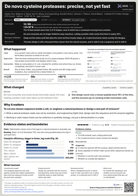
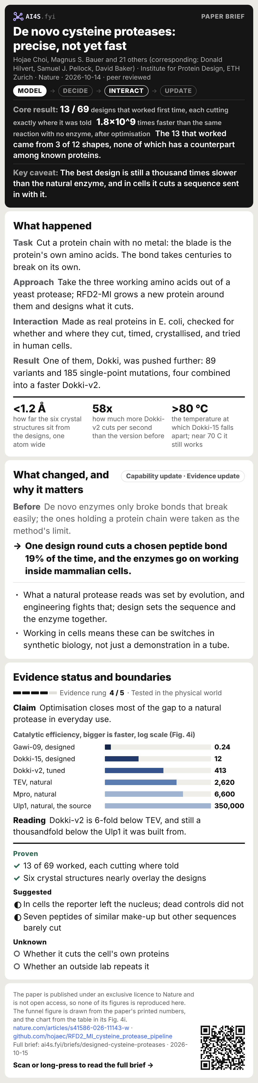
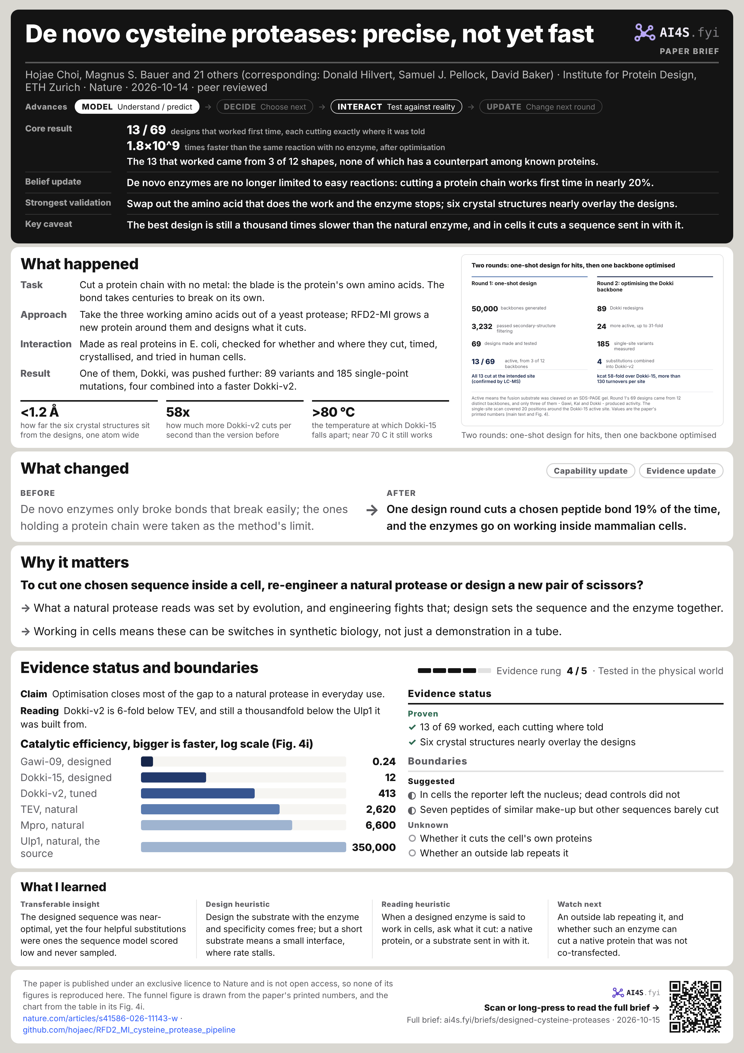
13 / 69designs that worked first time, each cutting exactly where it was told
1.8×10^9times faster than the same reaction with no enzyme, after optimisation
The 13 that worked came from 3 of 12 shapes, none of which has a counterpart among known proteins.
- Belief update
- De novo enzymes are no longer limited to easy reactions: cutting a protein chain works first time in nearly 20%.
- Strongest validation
- Swap out the amino acid that does the work and the enzyme stops; six crystal structures nearly overlay the designs.
- Key caveat
- The best design is still a thousand times slower than the natural enzyme, and in cells it cuts a sequence sent in with it.
01What happened
- Task
- Cut a protein chain with no metal: the blade is the protein's own amino acids. The bond takes centuries to break on its own.
- Approach
- Take the three working amino acids out of a yeast protease; RFD2-MI grows a new protein around them and designs what it cuts.
- Interaction
- Made as real proteins in E. coli, checked for whether and where they cut, timed, crystallised, and tried in human cells.
- Result
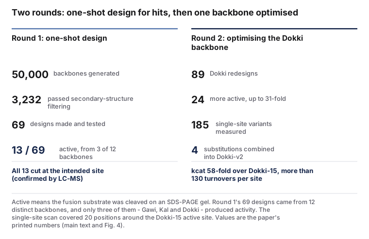
- One of them, Dokki, was pushed further: 89 variants and 185 single-point mutations, four combined into a faster Dokki-v2.
<1.2 Åhow far the six crystal structures sit from the designs, one atom wide
58xhow much more Dokki-v2 cuts per second than the version before
>80 °Cthe temperature at which Dokki-15 falls apart; near 70 C it still works
On 14 October 2026 Nature published two enzyme-design papers from the Institute for Protein Design on the same day; this is the cysteine-protease one (s41586-026-11143-w, received 14 October 2025, accepted 9 September 2026). The first authors are Hojae Choi and Magnus S. Bauer, the corresponding authors Donald Hilvert at ETH Zurich and Samuel J. Pellock and David Baker at the University of Washington, with 23 authors in all. The authors declare no competing interests; the design pipeline and analysis code are on GitHub and the six crystal structures are in the PDB (9YNL, 9YNM, 9YOX, 9YOY, 9YOZ, 9YP0). Every number on this page was read in the paper's own text and figure captions, from the PDF the author supplied.[S1][S2]
The pipeline: move Ulp1's active site into a new backbone, and design the substrate with it
The motif comes from the crystal structure of the yeast SUMO protease Ulp1 (PDB 1EUV): the catalytic triad Cys580, His514 and Asp531, an oxyanion hole formed by the Gln574 side chain and the 578-579 backbone, and four residues of substrate. A cysteine suits this job better than a serine: its thiol is more readily deprotonated with the histidine's help, and the high-energy intermediates it forms are more stable. Where the substrate sits is the other half of the problem: the authors had it pair with the enzyme as a beta strand, which is how Ulp1 itself binds, using backbone hydrogen bonds to hold the scissile amide beside the catalytic machinery. RFD2-MI, the molecular-interface version of RFdiffusion2, generates a 180-200 residue protein around that motif with a 7-residue substrate segment; what matters is that it conditions on where the catalytic atoms are without having to say which sequence positions carry them. Of 50,000 initial backbones, secondary-structure filtering (dropping long loops, low strand content, high coil) left 3,232, and several rounds of AlphaFold 3 scoring and partial diffusion followed; sequences came from ProteinMPNN, with enzyme and substrate optimised together, so each protease carries a substrate sequence of its own.[S1]
Screening was made cheap: the protease and its substrate sit on one chain, followed by mScarlet and a His tag, so an active design cuts itself during expression in E. coli and the molecular-weight shift shows on a gel after purification. Of 69 designs, 13 cut (19%), from 3 of 12 distinct backbones, which the authors named Gawi, Kal and Dokki. Dokki and Kal cleaved completely, Gawi about 95%. LC-MS confirmed that all 13 cut precisely at the intended site. These 13 are not yet usable enzymes: moved to a FRET assay with protease and substrate apart, the Gawi family was too weak for kinetics and the Dokki family's kcat sat around 10^-4 s^-1. So the second round was optimisation rather than more designs: 21 redesigns for Gawi and 89 for Dokki, of which 7 and 24 were more active, by 1.2- to 31-fold.[S1]
02What changed
Capability updateEvidence updateDe novo enzymes only broke bonds that break easily; the ones holding a protein chain were taken as the method's limit.
One design round cuts a chosen peptide bond 19% of the time, and the enzymes go on working inside mammalian cells.
What changed is which reactions are reachable, and where the result can be used. The paper opens by naming the difficulty precisely: resonance makes an amide far harder to break than an ester, the amine leaving group in a protein has a deprotonation pKa above 35 against under 8 for a typical activated ester, and peptide substrates are flexible and hard to bind precisely. Earlier de novo hydrolases sidestepped the first two. This work takes all three together and gets a 19% one-shot hit rate: high for de novo enzyme design, and the authors call it a relatively high success rate themselves. Something else changed too: a Foldseek search put the designs' closest match to any known protein at a TM score of 0.46, where above 0.5 indicates structural similarity, and structural alignment of even those top hits showed clearly distinct architectures with backbone RMSDs of several angstroms. These are not natural proteins with a few residues swapped; they are working enzymes in parts of structure space nature has not occupied.[S1]
03Why it matters
To cut one chosen sequence inside a cell, re-engineer a natural protease or design a new pair of scissors?
- What a natural protease reads was set by evolution, and engineering fights that; design sets the sequence and the enzyme together.
- Working in cells means these can be switches in synthetic biology, not just a demonstration in a tube.
The cell experiment is the part worth reading carefully, because it is both a real result and an easy one to overstate. The setup: a reporter, H2B-sfGFP-substrate-mScarlet, is transfected into HeLa cells, where H2B's nuclear localisation signal tethers the whole fusion in the nucleus, and a designed protease is co-transfected. If the enzyme cuts the substrate in the middle, mScarlet is released and redistributes out of the nucleus, visible as the red and green signals separating. Dokki, Dokki-15 and Dokki-v2 all did it, while Cys-to-Ala knockout controls kept both signals colocalised in the nucleus. Three independent transfections, nine fields each, about 200 cells analysed per condition. So what can be said is that these enzymes fold, stay active and cut their designed sequence in a mammalian cell; what cannot is that they cut any endogenous protein, since the substrate was designed by the authors and transfected in with them.[S1]
The specificity side is just as concrete. Dokki-15 strongly prefers its own substrate, VPTLTGG/G, and shows minimal or no detectable cleavage of seven non-cognate peptides of similar amino-acid composition. The authors point out that several of those also carry hydrophobic residues in the P4-P6 positions and still were not cut efficiently, so what is being read is the sequence, not just a hydrophobic pattern. The consequence for use is direct: a protease that recognises one roughly ten-residue sequence is the shape an orthogonal switch in synthetic biology needs, cutting a linker someone designed and leaving the rest alone. Seven non-cognate peptides is what that rests on, not a proteome-wide test, which is the experiment that would settle it.[S1]
04Evidence status and boundaries
Evidence rung 4 / 5 · Tested in the physical world · Physical validationEvidence status
Proven
- 13 of 69 worked, each cutting where told
- Six crystal structures nearly overlay the designs
Boundaries
Suggested
- In cells the reporter left the nucleus; dead controls did not
- Seven peptides of similar make-up but other sequences barely cut
Unknown
- Whether it cuts the cell's own proteins
- Whether an outside lab repeats it
ClaimOptimisation closes most of the gap to a natural protease in everyday use.
Catalytic efficiency, bigger is faster, log scale (Fig. 4i)
ReadingDokki-v2 is 6-fold below TEV, and still a thousandfold below the Ulp1 it was built from.
The catalysis really is the designed triad.
For each active design the authors mutated the catalytic cysteine, the histidine and the oxyanion-hole glutamine or asparagine to alanine one at a time. Replacing the Cys or the His abolished activity, confirming that neither is replaceable; replacing the Gln or Asn did not uniformly inactivate the designs, which the authors suggest may mean ordered water molecules can partly take over stabilising the oxyanion intermediate in its absence. Six crystal structures, at resolutions from 1.56 to 3.38 A, cover apo and substrate-bound states across three backbones: in the Kal-substrate complex both the triad and the substrate-binding cleft adopt the designed conformations; in apo Gawi-09 the catalytic residues align with the model except for a histidine displaced by a zinc ion from the crystallisation buffer; and in apo Dokki and Dokki-15 the density shows the catalytic cysteine oxidised to a sulfonic acid, which has also been reported for the natural cysteine protease Ulp1.[S1]
The rate: measured against what, and how far short.
Steady-state kinetics for two representative designs: Gawi-09 at kcat 0.8+/-0.1x10^-5 s^-1, KM 34+/-4 uM and kcat/KM 0.24+/-0.03 M^-1s^-1; Dokki-15 at kcat 1.1+/-0.1x10^-3 s^-1, KM 89+/-10 uM and kcat/KM 12+/-2 M^-1s^-1. Against the uncatalysed rate of peptide hydrolysis (3.6x10^-11 s^-1 at 25 C and pH 6.8), Dokki-15's acceleration is about 3x10^7-fold, which is the figure in the abstract. The authors then made 185 single-site variants at 20 positions around the Dokki-15 active site: most substitutions reduced activity, indicating the designed sequence was already near-optimal, but Tyr41, Ser44, Pro67 and Thr99 were beneficial, and all four had been given low probabilities by ProteinMPNN and never sampled in the original design. The resulting Dokki-v2 has kcat 0.064+/-0.005 s^-1, KM 154+/-14 uM and kcat/KM 413+/-50 M^-1s^-1, over 130 turnovers per active site, and a 1.8x10^9-fold acceleration; the whole gain comes from a 58-fold rise in kcat, meaning the chemical step itself got faster. For comparison: Ulp1 on its SUMO-containing substrate runs at kcat 6.1 s^-1 and kcat/KM 350,000; TEV at 0.16 and 2,620; Mpro at 1.23 and 6,600. Dokki-v2 sits at the lower edge of the natural range.[S1]
Why the gap remains.
The authors do not leave the remaining gap as a promise to improve; they localise it. PLACER ensembles of the catalytic cycle give the fraction of productive conformations, judged by geometry thresholds taken from natural cysteine proteases (Cys-His under 4.2 A, His-Asp under 3.2 A, oxyanion-Gln under 3.4 A): at the first transition state Ulp1 is at 100%, Dokki at 12%, Dokki-15 at 20% and Dokki-v2 at 32%. Molecular dynamics gives a second reason, and it is about the substrate rather than the enzyme: Ulp1 grips its substrate with a folded SUMO domain that pushes the scissile bond into the active site, while these designs read a short P1-P7 peptide across a smaller interface with fewer hydrophobic contacts, so the substrate is less firmly positioned. One telling control: Ulp1 itself shows no detectable activity on peptide substrates that lack the SUMO domain. The authors' conclusion is therefore that the next steps are active-site preorganisation and a larger substrate-binding interface, not different catalytic chemistry.[S1]
05What I learned
- Transferable insight
- The designed sequence was near-optimal, yet the four helpful substitutions were ones the sequence model scored low and never sampled.
- Design heuristic
- Design the substrate with the enzyme and specificity comes free; but a short substrate means a small interface, where rate stalls.
- Reading heuristic
- When a designed enzyme is said to work in cells, ask what it cut: a native protein, or a substrate sent in with it.
- Watch next
- An outside lab repeating it, and whether such an enzyme can cut a native protein that was not co-transfected.
Two papers on one day: double evidence that a capability has arrived
Nature published this institute's metalloprotease paper the same day (this site has a brief on it too): the same hard reaction, cutting a peptide bond, through entirely different chemistry (a zinc-activated water rather than an activated cysteine) with 135 designs in one round and 36% active. Two routes that do not depend on each other, delivered on the same day, look less like one method working and more like the design capability itself crossing a threshold. Both papers diagnose the remaining gap in the same place: incomplete preorganisation, with neither the catalytic groups nor the substrate held where they should be. What to keep in mind is that both come from the same lab system, so they corroborate each other rather than replicate each other.
A gap someone put a number on
The PLACER numbers deserve recording on their own: the fraction of productive conformations is 100% for Ulp1, 12% for Dokki, 20% for Dokki-15 and 32% for Dokki-v2. They turn 'why is a designed enzyme slower than a natural one' from a qualitative remark into a quantity that can be tracked, and one that orders the designs the same way their measured kcat does. For anyone doing generative design this points at a more useful metric than hit rate: a structure predictor's confidence says whether the fold is right, while catalysis needs to know how much of the time the active site actually sits where the reaction requires. The first is now done well; the second is the work that remains.
As one imageTo save and share→Sources
Factual claims link to original announcements, project lists, trial records or journal papers where possible. Research plans are kept separate from completed results.
- S1De novo design of cysteine proteasesNature · Choi, Bauer, Hilvert, Pellock, Baker et al. · Institute for Protein Design, ETH Zurich · 2026.10.14 · Peer-reviewed paper · full text and figures read from the PDF the author supplied, 2026-10-15
- S2hojaec/RFD2_MI_cysteine_protease_pipelineGitHub · named in the paper's code-availability statement · 2026.10.14 · The design and analysis code, from the paper's code-availability statement